As yet there is no effective treatment or cure for inherited retinal diseases. The great hope is that scientific research will unlock the knowledge of how to effectively treat and prevent blindness caused by these degenerative diseases.
What research are we funding?
We are currently helping to fund two research projects…
ONE
The Australian IRD Register and DNA Bank.
This vital Perth based resource contains data from over 6,200 Australian IRD families, and includes DNA samples from nearly 5000 of them.
The plan is to continue to expand the database, and collect DNA from more affected families, and to analyse as many samples as possible.
This is a world-leading source of data, which is being used for international clinical trials into IRD.
We have been funding this project since 1984, and have committed to giving a further $50,000 per year until at least 2018.
TWO
Accelerating Therapeutic Discoveries for Retinitis Pigmentosa.
This project is run by the Lions Eye Institute of WA and the Vision Regeneration Laboratory Centre for Eye Research Australia in Victoria.
Its aim is to successfully grow stem cells from RP affected individuals. These cells will be further investigated, and used to trial new treatments.
This research has already resulted in several major discoveries. The ultimate goal is prevention of blindness, and restoration of sight to people affected with RP.
Researchers are also investigating a number of other avenues to prevent the process of premature cell destruction in the retina.
Although it is not possible to transplant the retina (because of its connection through the optic nerve to the brain), transplantation of cells nourishing the retina promises in the near future to become a practical technique for preserving and perhaps improving sight.
These exciting prospects opening up through research are made possible by generous public donations around the world. But research is very expensive and what can be achieved is restricted by the money available.
Retina Australia (WA) finances research in Australia for inherited retinal diseases and has an urgent need of funds for this purpose.
Donations can be made online through Givematcher.com – note all proceeds rasised through Givematcher come directly to Retina Australia WA as they change 0% fees or commissions.

****************************************
RESEARCH FUNDED BY RETINA AUSTRALIA 2013
Introducing Biorad at Lions Eye Institute – Funding provided by Retina Australia WA Inc in July 2013
2 October 2013 · ![]() · Taken at Lions Eye Institute
· Taken at Lions Eye Institute

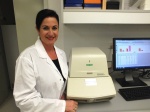
Retina Australia (WA) are proud to announce the donation of a Real time PCR machine (Biorad CFX CON NECT SYSTEM) to the Lions Eye Institute (LEI) new facility, which is due to open late this year. The CFX Connect real-time PCR detection system offers two-target analysis, excellent thermal cycler specifications, and incorporates innovative optical technologies with powerful software to provide maximal reliability and efficiency for all real-time PCR needs.
****************************************
RESEARCH FUNDED BY RETINA AUSTRALIA 2012
“Chen & Mackey” | University of Western Australia | Project – ‘Microperimetry natural history study of Stargardt’s macular dystrophy.
Stargardt disease is the most common hereditary macular dystrophy, occurring in 1 in 10,000 population. This incurable condition presents with bilateral central visual loss in the first few decades of life which may progress to legal blindness over many years or decades in up to 50% of affected individuals. Early progression of the macular degeneration is slow and often undetectable using routine vision tests in the short-term. Visual acuity reduction may take several years or decades due to sparing of the fovea even in advance stages of the disease. This unique feature of Stargardt’s disease poses a special challenge for designing clinical trials which aim to demonstrate efficacy within 1-2 years. This is because regulatory authorities still insist that visual acuity is used as a primary endpoint.
Therefore, there is an urgent need to find and validate an alternative treatment endpoint which has the ability to demonstrate progression or stabilisation of Stargardt’s disease within a 6-12 months time frame so that the effectiveness of novel therapies can be detected earlier.
In this study, we will specifically examine the natural history of patients with Stargardt’s disease so that we can determine if there’s significant decline in retinal function in these patients over a 12 month period. Visual acuity, microperimetry, (a specialised visual field test) and retinal photography will be performed on both eyes of each patient at baseline, 2, 4, 6, 9 and 12 months. Results will be useful for planning future treatment trials in Stargardt’s disease.
The study will draw on Dr Chen’s previous work which demonstrated that microperimetry can detect significant decline in retinal function adjacent to areas of atrophy in nine patients. Amongst these, three had Stargardt’s disease.
“Valter & Madigan” | The Australian National University and the University of NSW | Project – ‘In vitro and In vivo response of retinal Müller glia to photobiomodulation’.
Müller glial cells extend across the retina, interacting with retinal neurons and blood vessels, and play a significant role in maintaining normal retinal function. When the retina is stressed, for example following bright light exposure, or when cells such as retinal photoreceptors degenerate, Müller cells become activated (reactive gliosis). Reactive gliosis is generally evidenced by increased cell proliferation, abnormal branching of cell processes, upregulation of structural proteins, cell migration and/or cell dedifferentiation.
Gliosis can induce either neuroprotective or neurodegenerative effects related to the degree and type of retinal stress. Neurotrophic factors released by glial cells may promote the survival of remaining neurons, however when Müller cells proliferate and grow into the subretinal space through breaks in the outer limiting membrane and in areas where there
is photoreceptor loss, gliosis and scar formation can inhibit or limit the potential for retinal remodelling and regeneration. Photobiomodulation is a non-invasive therapeutic approach where light of a certain wavelength can induce a cellular response that promotes tissue regeneration. Near infrared light (NIR), specifically, light at 670nm wavelength, is known to
ameliorate light-induced loss of retinal photoreceptors and reduce retinal inflammation.
However its effects on Müller glial activation and gliosis are not well understood. In this project we will investigate the effects of NIR on Müller cells in culture, and also in a model of retinal degeneration (light-induced retinal damage).
********************************************
RESEARCH FUNDED BY RETINA AUSTRALIA 2011
The promising research by Dr Fletcher’s team has continued to be funded.
The University of Sydney | Save Sight Institute
Children’s Hospital Westmead, NSW and Children’s Medical Research Institute
“Investigation for novel disease genes in cone and rod dystrophies”
Chief Investigators: Dr Robyn Jamieson and Dr John Grigg
The University of Western Australia
“Role of a novel miRNA in the dominant syndromic disorder of macula dystrophy and split hand and foot malformation”
Chief Investigators: Professor David M Hunt and Professor Shaun P Collin.
Centre for Eye Research Australia and University of Melbourne Departmentof Ophthalmology Royal Victorian Eye and Ear Hospital
“Does the level of systemic inflammation influence the outcome of treatment in neovascular Age-Related Macular Degeneration”
Chief Investigators: Professor Robyn Guymer, and Dr Lyndell Lim
********************************************
RESEARCH FUNDED BY RETINA AUSTRALIA 2010
Report from Dr Ursula Grefarath, Melbourne University
Histamine in the development and maintenance of the retina

The second grant of $23,600 was awarded to Dr Erica Fletcher and her team whose research is also in search of a treatment. “Understanding how photoreceptors die is crucial for the development of therapeutic agents that slow the progression of RP. The research is aimed at examining in detail how the molecule, ATP, causes photoreceptor death, and whether blockade of this class of molecule slows photoreceptor death.”
This funding is the result of a significant fund-raising effort by RA supporters, especially in view of the economic downturn experienced by the community in 2009.
It is also over and above the ongoing cost of our national Inherited Retinal Disease Register and DNA Bank for families in Australia affected by Retinitis Pigmentosa and other Retinal Dystrophies.
**************************************
RESEARCH FUNDED BY RETINA AUSTRALIA 2008
Report from professor Michael Kalloniatis, University Of Auckland
photoreceptor degeneration in Retinitis Pigmentosa
Report from Dr Erica Fletcher , University of Melbourne
The role of purines in photoreceptor death during retinal degeneration
**************************************
RESEARCH FUNDED BY RETINA AUSTRALIA 2006
Report from Professor Michael Kalloniatis, University of Auckland.
Photoreceptor degeneration in Retinitis Pigmentosa
Report from Professor P E Rakoczy from the Lion’s Eye Institute, WA
Optimising the benefits of retinal gene therapy using animal models of Leber’s Congenital Amaurosis
Report from Dr Nigel Lovell, University of New South Wales
***********************************
RESEARCH FUNDED BY RETINA AUSTRALIA 2005
